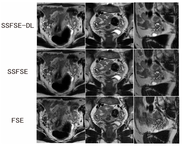
Figure 2.
Ovarian MRI in a 28-year-old woman with confirmed PCOS. SSFSE-DL images (upper row) show the least noise and blurring artifacts. The bilateral enlarged ovaries with many small peripheral follicles are clearly delineated on the SSFSE-DL images. The display of the follicles is impaired by the noise on the SSFSE images (middle row) and by the blurring artifacts in the FSE images (lower row).

