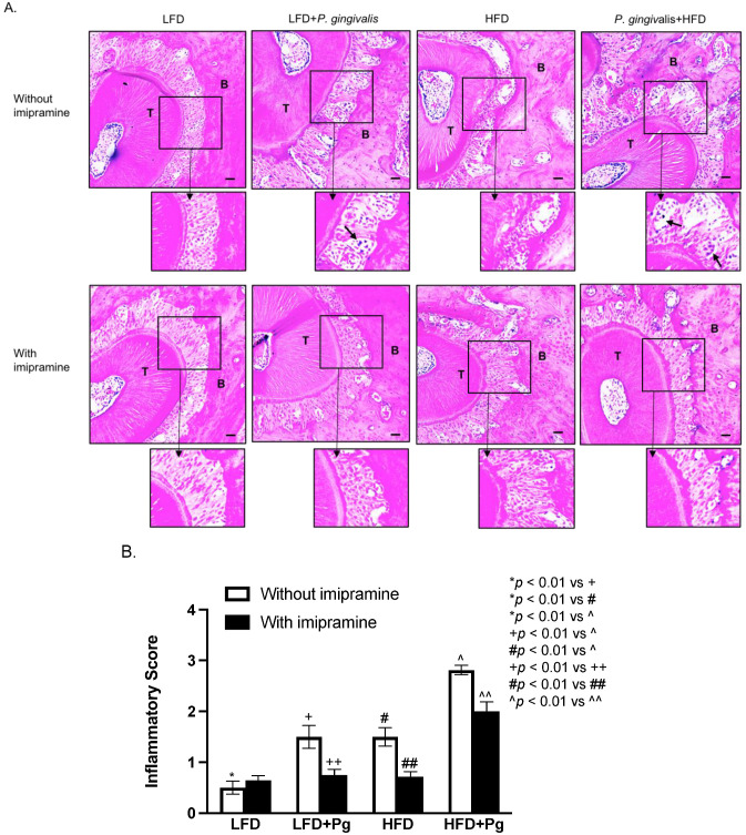
Figure 4.
Histological analysis of periodontal tissues of mice after HFD feeding, oral P. gingivalis inoculation, and imipramine treatment. After performing the mCT of the maxillae as described above, the tissues were sectioned and stained with hematoxylin and eosin (H/E) for histological evaluation of leukocyte infiltration and bone resorption. (A) The images of the representative tissue sections with H/E staining focus on the area of the periodontal ligament, teeth, and alveolar bones. The images in the insets were enlarged, and the arrows indicated infiltrated leukocytes. Scale bar = 100 μm. (B) The inflammatory scores were made according to the degree of leukocyte infiltration and bone resorption as described in Materials and Methods. The data are mean ± SD (n = 12). T: tooth; B: alveolar bone.

