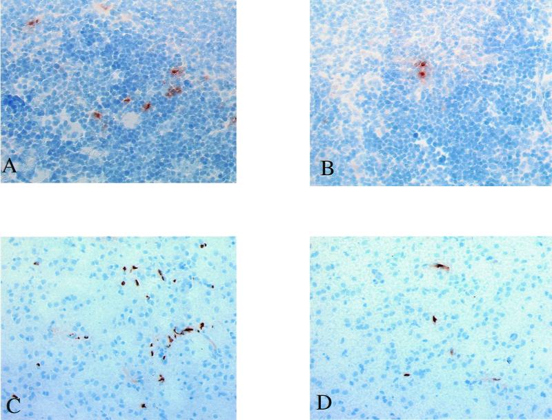
FIG. 3.
(A and B) Microphotographs of cryopreserved spleen sections immunohistochemically stained for TNF-α 3 h after Hib inoculation. Sections from an untreated control animal (A) and a CNI-1493 treated animal, where the number of positively stained cells was reduced (B), are shown. TNF-α-expressing cells stain brown with diaminobenzidine. Please note the extracellular deposition of TNF surrounding producer cells. The nuclei of all cells were counterstained with hematoxylin (blue). (C and D) Microphotographs of cryopreserved brain sections immunohistochemically stained for infiltrating granulocytes 24 h after Hib inoculation. Sections from an untreated control animal (C) and a CNI-1493-treated animal, showing a reduced number of granulocytes (D), are shown. Granulocytes stained brown with diaminobenzidine. The nuclei of all cells were counterstained with hematoxylin (blue).

