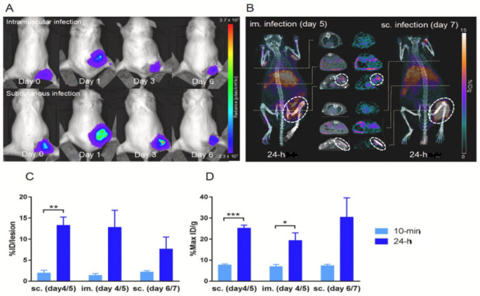
Figure 3.

64Cu-loaded liposome PET/CT of a S. aureus Xen29 infectious mouse model. (A) BLI of female NMRI mice sc. or im. injected with S. aureus Xen29. Mice were scanned on day 0, 1, 3, and 6 after bacterial inoculation. (B) PET/CT images of S. aureus Xen29 infection 24 h after iv. injection with 64Cu-liposomes. White rings encircle lesion sites and 64Cu-liposome activity. (C,D) Activity levels of 64Cu-liposomes on PET/CT scans performed on day 4 and 5 (acute) and on day 6 and 7 (chronic) after bacterial inoculation. All mice were scanned 10 min and 24 h after injection with 64Cu-liposomes. Data are presented as mean ± SEM. Significant differences presented as * p < 0.05; ** p < 0.01; *** p < 0.001; paired t-test. sc.: subcutaneous; im.: intramuscular. Reprinted with permission from Ref. [127]. Copyright 2020 Elsevier.
