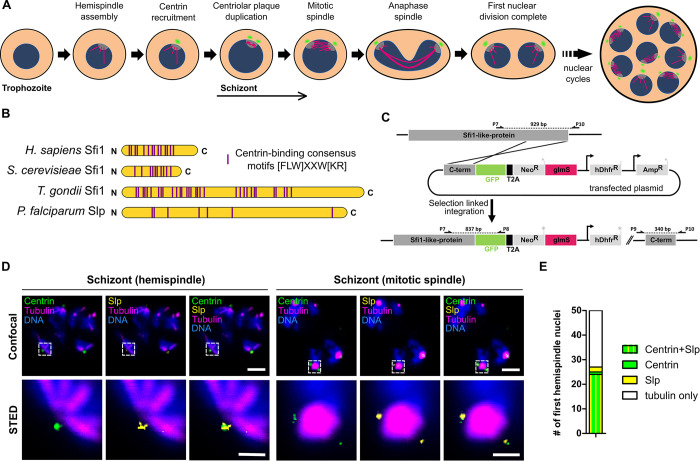
Fig 1. The Sfi1-like protein (PfSlp) co-localizes with centrin at the centriolar plaque.
A) Schematic of first nuclear division in Plasmodium falciparum during blood stage schizogony (red blood cell not shown). After polymerization of hemispindle microtubules (magenta) at the inner core of the CP (grey), centrin (green) is recruited to the outer core of the CP. The centriolar plaque is duplicated, and the mitotic spindle is formed. During anaphase chromosomes (blue) are segregated and two nuclei are formed. Subsequent asynchronous nuclear division cycles lead to a multinucleated cell stage, which later gives rise to multiple daughter cells. B) Schematic comparison of human, Saccharomyces cerevisiae, and Toxoplasma gondii Sfi1 with PfSlp. No clear sequence homology to other Sfi1 proteins exists, but multiple centrin-binding site consensus motifs can be identified. C) Endogenous PfSlp tagging strategy with GFP and glmS ribozyme via selection-linked integration (SLI). D) Confocal microscopy images of immunofluorescence staining of blood stage schizont expressing endogenously tagged PfSlp-GFP using anti-PfCen3, which labels multiple centrins (green), anti-tubulin (magenta), anti-GFP (yellow) antibodies, and DNA stained with Hoechst (blue). Dual-color STED microscopy of marked sub-region (dotted rectangle) with super-resolved PfSlp-GFP and centrin signal. Maximum intensity projections are shown for confocal images. Scale bars, confocal, 1.5 μm; STED, 0.5 μm. E) Quantitative analysis of PfSlp, centrin and tubulin immunostained parasites in mononucleated schizonts with hemispindle. CPs were scored for presence (27/50) and absence (23/50) of centrin and/or PfSlp signal.

