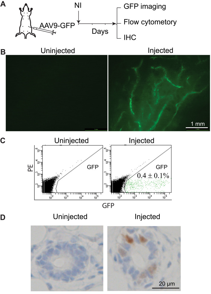
Fig. 1. AAV serotype 9 infects mammary gland epithelial cells.
(A) Diagram of experimental design to test the infection capacity of AAV serotype 9. (B) Mammary duct tree 3 days after intraductal infection by AAV9 carrying copGFP, imaged under a fluorescent stereoscope. (C) Detection of AAV9-GFP–infected cells using flow cytometry. PE, phycoerythrin. (D) Detection of AAV9-GFP–infected cells using immunohistochemistry (IHC).

