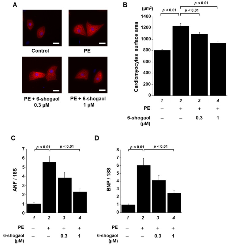
Figure 2.
6-shogaol treatment significantly suppressed PE-induced cardiomyocyte hypertrophy in vitro. Primary cultured cardiomyocytes derived from neonatal SD rats were pretreated with 6-shogaol (0.3 or 1 μM) and then treated with the stimulant PE (30 μM) for 48 h. (A) Immunofluorescence staining was then carried out using anti-MHC antibody and Alexa Fluor 555-conjugated anti-mouse IgG. Scale bar: 20 μm. (B) ImageJ software was used to measure cell surface area (50 cells/well). The mean ± SEM values of three independent experiments are shown by the bars. The mRNA levels of hypertrophy-related ANF (C) and BNP (D) gene transcriptions were examined by quantitative RT-PCR. The mean ± SEM of four individual experiments is shown by the bars.

