
Figure 1. NK cells accumulate in skin wounds.
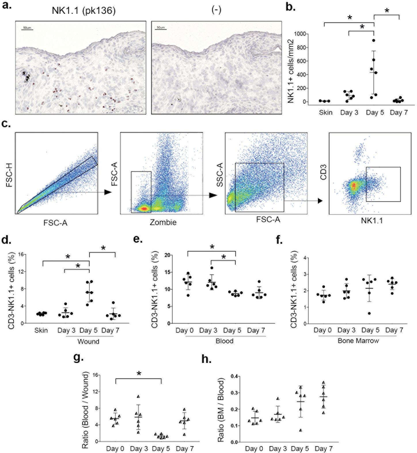
(a) NK cells identified in cryosections by immunohistochemical staining for NK1.1 (pk136) in male mice and (b) corresponding summary data for cell counts. (c) NK cells also assessed by flow cytometry: gating strategy shown for identifying NK cells (CD3−NK1.1+), Zombie dye used for gating viable cells from wounds. (d) Summary data for NK cells in wounds, (e) blood and (f) bone marrow. (g) Ratio of NK cells in blood/wound and (h) in bone marrow/blood. Data presented as mean ± SD, n = 6 mice, from two separate experiments in cohorts of 3 mice each. For each data set, one-way ANOVA and Tukey’s post hoc test were performed. *p < 0.05 between indicated time points.