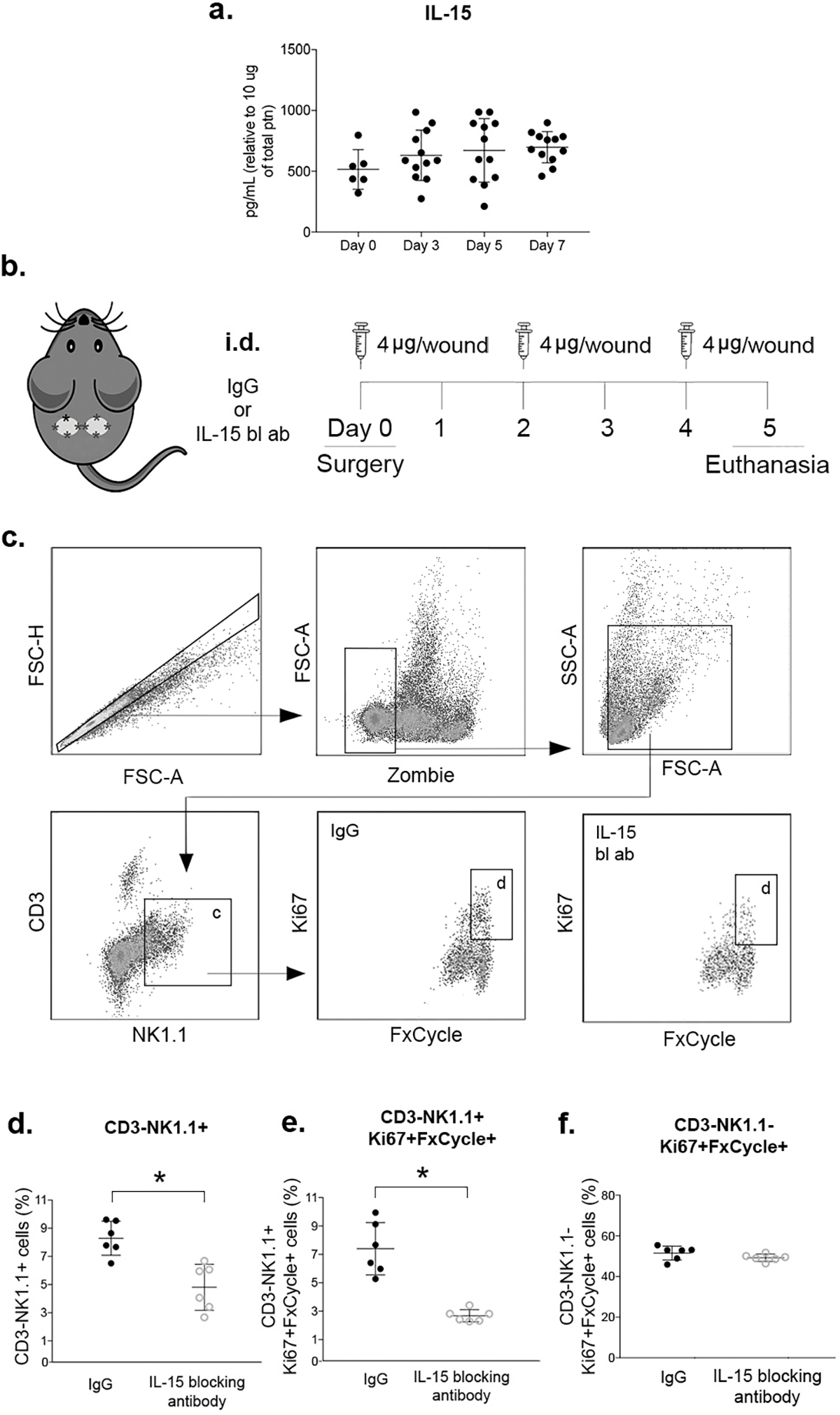
Figure 2. NK cells proliferate in wounds in an IL-15-dependent manner.

(a) Levels of IL-15 protein in wounds measured by flow cytometry multiplex assay. (b) Local IL-15 blocking strategy: animals were subjected to excisional wounding and then treated with intradermal (i.d.) injections of 4ug of anti-mIL-15 antibody or IgG isotype control antibody on days 0, 2 and 4. Wounds harvested on day 5. (c) Gating strategy for NK cells (CD3−NK1.1+) and proliferating NK cells (Ki67+FxCycle+) identification by flow cytometry, Zombie dye used for gating viable cells. (d) Summary data for NK cells (CD3−NK1.1+) in wounds. (e) Summary data for proliferating NK cells (CD3−NK1.1+Ki67+FxCycle+) in wounds. (f) Summary data for proliferating CD3−NK1.1− cells. Data presented as mean ± SD, n = 6 mice, from two separate experiments in cohorts of 3 mice. For IL-15 protein data, one-way ANOVA and Tukey’s post hoc test showed no significant differences. For IL-15 blocking antibody experiments, two-tailed t-tests showed significant differences between groups treated with anti-mIL-15 antibody and IgG isotype control;*p < 0.05.
