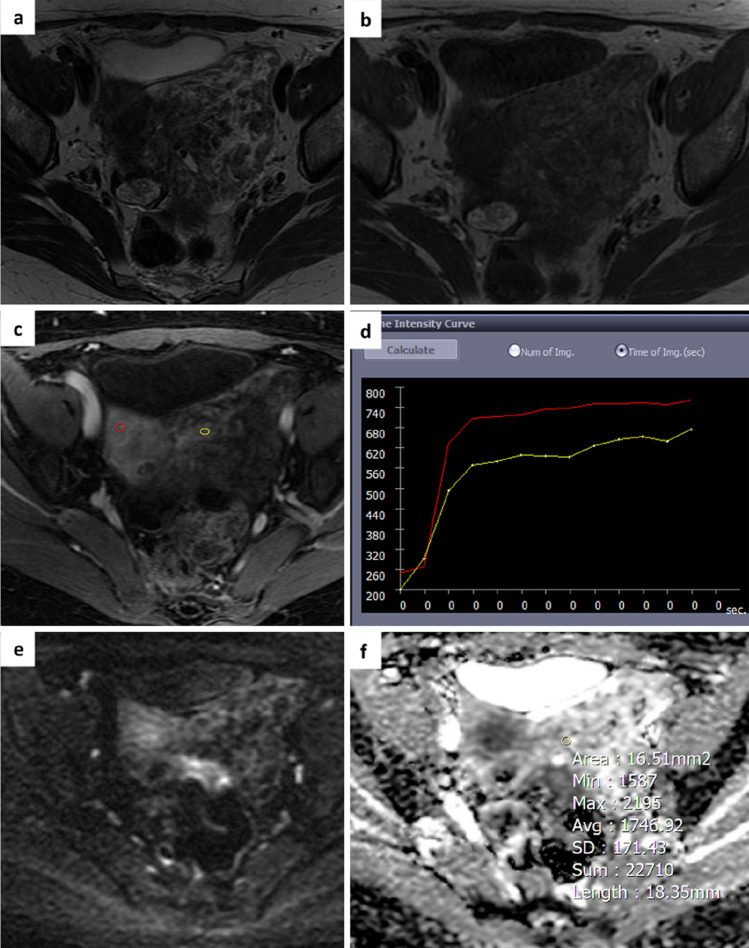
Fig. 3.
A 55-year-old woman with an indeterminate adnexal mass on ultrasound. Axial T2 and T1-weighted images show a voluminous mass in the left paramedian pelvis with mixed content due to the coexistence of solid components and fluid lacunae a,b. The perfusion sequence generates an intermediate risk curve (TIC type 2) c,d and an O-RADS MRI score 4 was attributed. DWI images and ADC map acquired in the axial plane show a slight restriction of the signal at b 1000 with an ADC value of 1.7 × 10⁻³ mm2/sec e,f. Histology revealed a benign lesion (ovarian fibroma)

