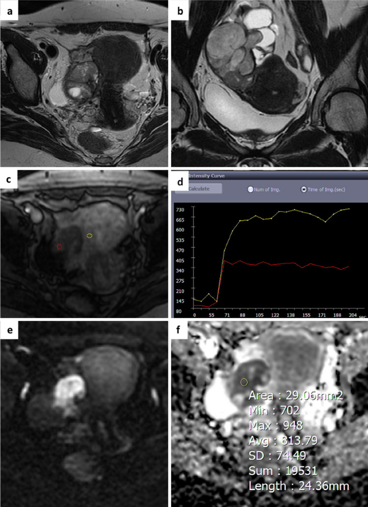
Fig. 4.
A 47-year-old woman. Axial and coronal T2-weighted images show a right adnexal mass with solid content and some fluid components a,b. The perfusion sequence generates an intermediate risk curve (TIC type 2) compared to the myometrium c,d and an O-RADS MRI score 4 was attributed DWI images and ADC map acquired in the axial plane shows high signal restriction at b 1000 with ADC value of 0.813 × 10⁻³ mm2/sec e,f. Histology revealed a malignant adnexal lesion (high-grade serous carcinoma).

