Figure 1.
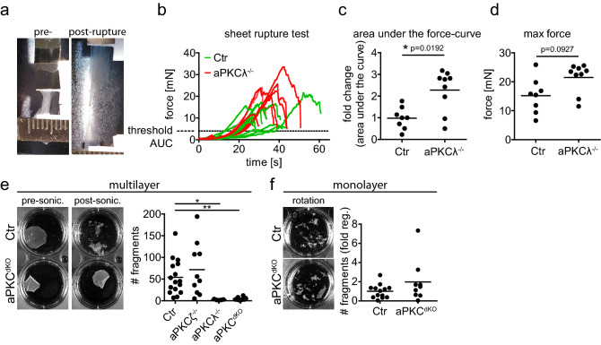
aPKC regulates mechanical resilience of stratified epithelial sheets. (a) Example of stratified keratinocyte sheet mounted in the tissue stretcher before (pre-) and after (post-) rupture. (b) Measured force over time while stretching with constant speed. (c) Quantification of the fold change of the area under the curve (AUC) from (b), normalized to the Ctr AUC mean value. With Mann–Whitney test for n = 8 (Ctr) and n = 9 (aPKCλ−/−). (d) Quantification of the maximum force that was reached during stretching, prior to sheet rupture. With Mann–Whitney test for n = 8 (Ctr) and n = 9 (aPKCλ−/−). Dots represent biological replicates. (e) Dispase assay with stratified keratinocyte multilayers (48 h Ca2+) after detachment upon dispase treatment. Images: Examples of sheets after end over end rotation, pre and post sonication. Graph: Quantification of sheet fragments upon sonication. *p < 0.05 with ANOVA followed by Dunnett’s multiple comparison test for n = 16 (Ctr), n = 5 (aPKCζ−/−), n = 10 (aPKCλ−/−), n = 10 (aPKCdKO). (f) Dispase assay with keratinocyte monolayers (6 h Ca2+) after detachment upon dispase treatment. Images: Examples of sheets after end over end rotation. Graph: Quantification of sheet fragments upon end over end rotation. n = 13 (Ctr), n = 9 (aPKCdKO).

