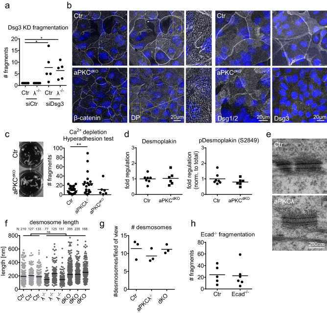
Figure 2.
Increased resistance does not depend on alterations in desmosomes. (a) Dispase assay with stratified keratinocyte multilayers (48 h Ca2+) transfected with either ctr siRNA (siCtr) or siRNA against desmoglein3 (siDsg3). Quantification of sheet fragments upon end over end rotation: *P = 0.0116 with 1-way ANOVA, Tukey’s post test for Ctr/aPKCλ−/−, siCtr/siDsg3: n = 5. (b) Immunofluorescence staining for β-catenin, desmoplakin (DP), desmoglein1/2 (Dsg1/2) and desmoglein3 (Dsg3) in stratified keratinocyte multilayers (48 h Ca2+). Enlargements show punctate desmosomal intercellular junctions. Nuclei labeled with DAPI (blue). (c) Quantification of sheet fragments upon EGTA treatment and end over end rotation. **P = 0.009 with 1-way ANOVA, Tukey´s post test for Ctr: n = 26, aPKCλ−/−: n = 19, aPKCdKO: n = 6. (d) Western Blot quantification of total and phospho-desmoplakin levels (Ser2849) in lysates of stratified keratinocyte sheets (48 h Ca2+). (e) Transmission electron micrographs, cross section of stratified keratinocyte sheets 48 h in high Ca2+ showing comparable desmosome in Ctr and aPKCλ−/− keratinocytes. Representative images from n = 3 (Ctr/aPKCλ−/−). (f, g) Quantification of desmosomal length (f) and quantity (g) from transmission electron micrographs as shown in (e) for Ctr/aPKCλ−/−/aPKCdKO: n = 3. Numbers of quantified desmosomes/biological replicate are indicated above the graph. (h) Dispase assay with stratified Ctr and E-cadherin−/− keratinocyte multilayers (48 h Ca2+) after detachment upon dispase treatment. Graph: Quantification of sheet fragments upon sonication. n = 5 (Ctr), n = 6 (E-cadherin−/−).

