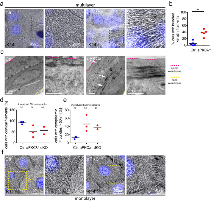
Figure 3.
aPKC regulates keratin filament organization. (a) Immunofluorescence analysis for keratin14 (K14) in apical cells of stratified keratinocyte cultures (48 h Ca2+). Nuclei labeled with DAPI (blue). (b) Quantification of cells with bundled keratin fibers. P = 0.0079, Mann–Whitney test, Ctr/aPKCλ−/−: n = 5. Dots represent biological replicates/mice. (c) Transmission electron micrographs, cross section of stratified keratinocyte sheets (48 h Ca2+) showing strong keratin bundles (white arrows) in suprabasal layers of aPKCλ−/− keratinocytes. Basal lamina is marked by the yellow dashed line. Representative images from n = 3 (Ctr/aPKCλ−/−). (d) Quantification of apical cells with cortical keratin filaments from EM images as shown in (c). (e) Quantification of apical cells with thick cytoplasmic keratin filaments from EM images as shown in (c). Dots represent biological replicates/mice. (f) Immunofluorescence analysis for K14 in keratinocyte monolayers (6 h Ca2+). Nuclei labeled with DAPI (blue). Representative example of > 3 biological replicates.

