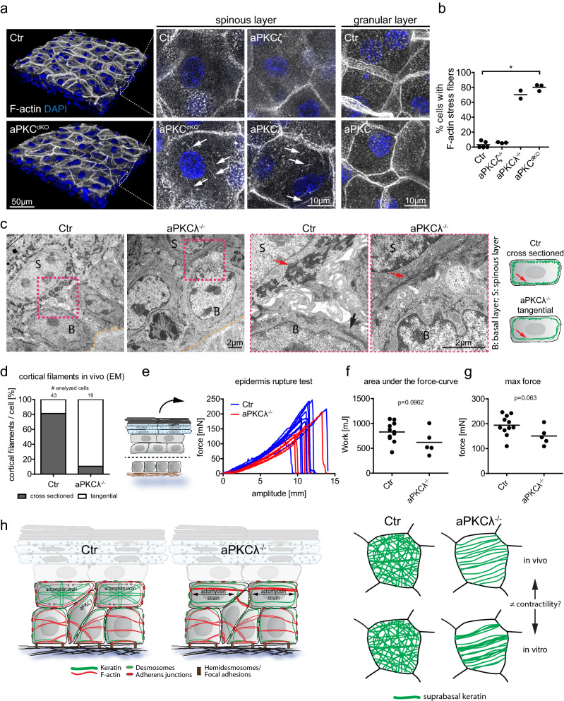
Figure 5.
aPKC regulates cytoskeletal reorganization and tissue resilience in vivo. (a) Newborn mouse epidermal whole-mount immunofluorescence analysis for F-actin revealing stress fibers (white arrows) in the first suprabasal layers upon epidermal loss of aPKCλ (aPKCλ−/−) or both aPKCλ and ζ (aPKCdKO) but not in Ctr or aPKCζ epidermal knockout (aPKCζ−/−) mice. (b) Quantification of cells with F-actin stress fibers in suprabasal (spinous) layers. Each dot represents one mouse. P = 0.0286 with Kruskal–Wallis, Dunn’s post hoc test for Ctr: n = 5, aPKCζ: n = 3, aPKCλ: n = 2, aPKCdKO: n = 3. (c) Transmission electron micrographs, cross section of newborn mouse epidermis showing an altered orientation of keratin bundles (red arrows) in suprabasal (spinous) layers of epidermal aPKCλ−/− mice. Basal lamina is marked by the yellow dashed line. Representative images from n = 4 (Ctr) and n = 3 (aPKCλ) mice. (d) Quantification of spinous cells with regular (cross sectioned) and irregular (tangentially cut or absent) keratin bundles. Cumulative values from n = 4 (Ctr) and n = 3 (aPKCλ) mice. (e) Measured force over time while stretching isolated suprabasal epidermis. (f) Quantification of the area under the curve from e. With Student´s t-test for n = 11 (Ctr) and n = 5 (aPKCλepi−/−). (g) Quantification of the maximum force that was reached during stretching, prior to sheet rupture. With Student’s t-test for n = 11 (Ctr) and n = 5 (aPKCλepi−/−). Dots represent biological replicates. (h) Model of aPKC dependent suprabasal intermediate filament organization.

