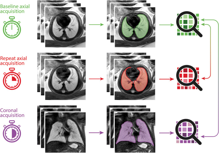
Fig. 1.
Graphical summary illustrating the segmentation and feature extraction process: A baseline axial T2-weighted sequence of the fetal lungs was acquired, and the lung manually segmented on all images. Three-dimensional lung masks were used to extract radiomics features from fetal MRI image data (top row, shown in green). This process was in a repeat axial acquisition (middle row, shown in red), and a coronal acquisition (bottom row, shown in purple). Lung radiomics features extracted from the baseline axial acquisition (shown in green) were compared to features extracted from the repeat axial (shown in red) and coronal (shown in purple) acquisitions to assess their reproducibility

