
Figure 2: Intrathecal AIBP reverses paclitaxel-induced allodynia and alters lipid rafts and TLR4 expression in DRG neurons.
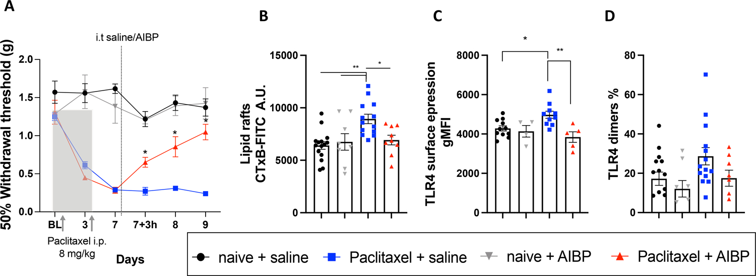
A. Hindpaw withdrawal thresholds in male mice in response to i.p. paclitaxel (2 injections of 8 mg/kg), followed by a single dose of i.t. saline (5 μl) or AIBP (0.5 μg/ 5 μl). Naïve mice received no i.p. injections (n=5–14 per group). Data shown from 2 independent experiments. B-D. Mouse groups shown in A were terminated on day 10, i.e, 72 hours after i.t. saline or AIBP, and DRG single-cell suspensions were analyzed by flow cytometry. Neurons were gated as CD45−/CD44−/CD24+ cells and analyzed for lipid raft content, measured by CTxB (lipid rafts) staining (B), total surface TLR4 (C); and dimerized TLR4 (D). Data shown from 2 independent experiments. Mean±SEM; *, p<0.05; **, p<0.01 (two-way (A) and one way (B-D) ANOVA).