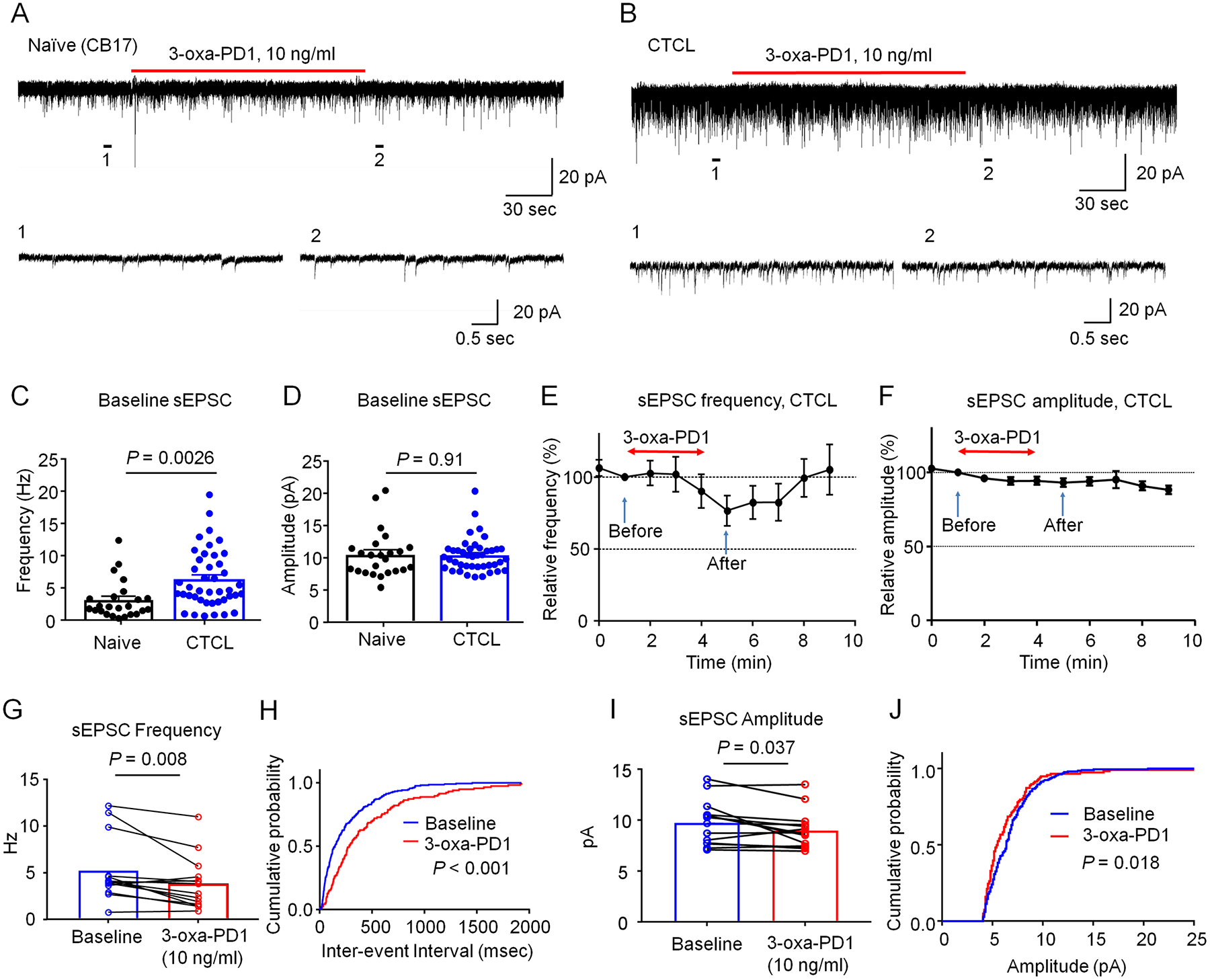
Figure 6. 3-oxa-PD1 inhibits excitatory synaptic transmissions in spinal cord slices of CTCL mice.

(A-B) Representative traces of spontaneous EPSCs (sEPSCs) in lamina IIo neurons of naïve CB17 (A) and CTCL (B) mice. Traces 1 and 2 are enlarged in the bottom panels. (C-D) Quantification of sEPSC frequency (C) and amplitude (D) in naïve and CTCL mice. CTCL significantly increased the frequency but not amplitude of sEPSC. Unpaired t-test, n = 24 neurons from 7 naïve mice and n = 42 neurons from 10 CTCL mice of both sexes. (E, F) Time course showing the effects of bath application of 3-oxa-PD1 (10 ng/ml, 3 min) on percentage changes of sEPSC frequency (E, n=14 neurons) and sEPSC amplitude (F, n=14 neurons). Arrows indicate before and after 3-oxa-PD1 perfusion. Note that 3-oxa-PD1 decreased the frequency but not amplitude of sEPSCs in spinal cord neurons of CTCL mice. Also note the effect of 3-oxa-PD1 can be washed out in 5 min. (G) sEPSC frequency. Paired t-test, n = 14 neurons from 5 CTCL mice. (H) Cumulative probability of inter-event interval shows longer interval after 3-oxa-PD1 application. Two-sample Kolmogorov-Smirnov test. (I) sEPSC amplitude. n = 14 neurons from 5 CTCL mice. (J) Cumulative probability of amplitude. The data were analyzed at 1 min (before treatment) and 5 min (after treatment). Data are shown as mean ± SEM.
