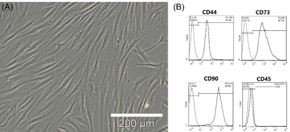
Fig. 1.

BMMSC Isolation and Identification. (A), BMMSCs (passage 3) were formed as densely packed spindle-shaped cells. The cells were isolated from two- to three-week-old rats (70-80 g). (B) Flowcytometric analysis was used to determine the cell surface markers of BMMSCs (passage3); high CD90 (99.9%), CD44 (99.9%), and CD73 (99.8%) expression was observed, whereas almost no CD45 expression was detected (1.25%).
