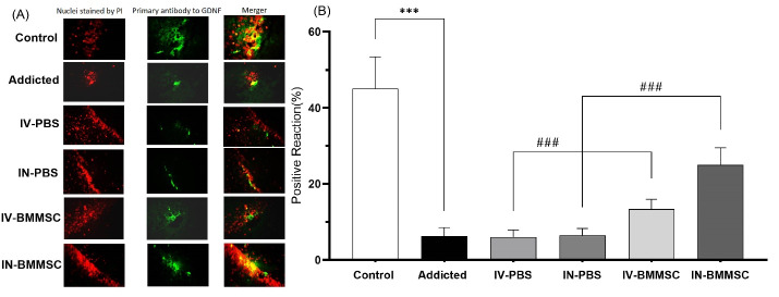
Fig. 7.
Distribution of immunoreaction products of GDNF in the hippocampus. GDNF protein was detected by Immunofluorescent in the hippocampus 2 weeks after the BMMSCs administration. (A) Nuclei staining by DAPI and primary antibody to GDNF and merge of them from each group (magnification, ×400). (B), The percentage of positive reaction in each group. ***P < 0.001 versus Meth-addicted group. ### P < 0.001 versus PBS-treated groups. Data are presented as the Mean ± SEM; Scale bar: 20 μm; GDNF, glia cell line derived neurotrophic factor. (C; Control group (n=8); PBS1, IV-PBS administration group (n=4); PBS2, IV-PBS administration group (n=4); A, Addicted group (n=7); T1, IV-BMMSCs administration group (n=8); T2, IN-BMMSC administration group (n=6).

