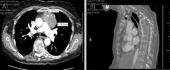
Figure 3.

Repeat computed tomography (CT) demonstrating mediastinal mass repeat (a) axial and (b) sagittal CT imaging demonstrating an irregular left anterior mediastinal mass measuring 5.7×3.5 cm with internal calcifications and left basilar compressive atelectasis. Also noted is a wedge compression deformity of the T7 vertebral body with retropulsion and subsequent spinal canal and left neural foraminal stenosis. There is also an associated soft-tissue mass eroding through the vertebral body and involving the intervertebral disk and superior endplate of the T8 vertebral body.
