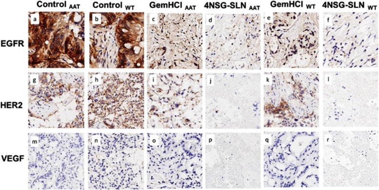
Fig. 9.
Expression of EGFR, HER2, and VEGFR receptors in GemHCl and 4NSG treated mice bearing PDX PCa tumor tissues from Black and White PCa patients. Representative images of 4NSG-SLNBT and 4NSG-SLNWT staining showed moderate EGFR expression (d and f) but negative staining for HER2 and VEGFR (j, l, p, and r). GemHClBT and GemHClWT revealed moderate to high EGFR, HER2, and VEGFR expressions (c, e, I, k, o, q) with 40X magnification. Representative images (a,g, and m) represented control AAT receptor expressions for EGFR, HER2, and VEGF, respectively, and images (b,h.n) represented control WT receptor expressions for EGFR, HER2, and VEGF, respectively

