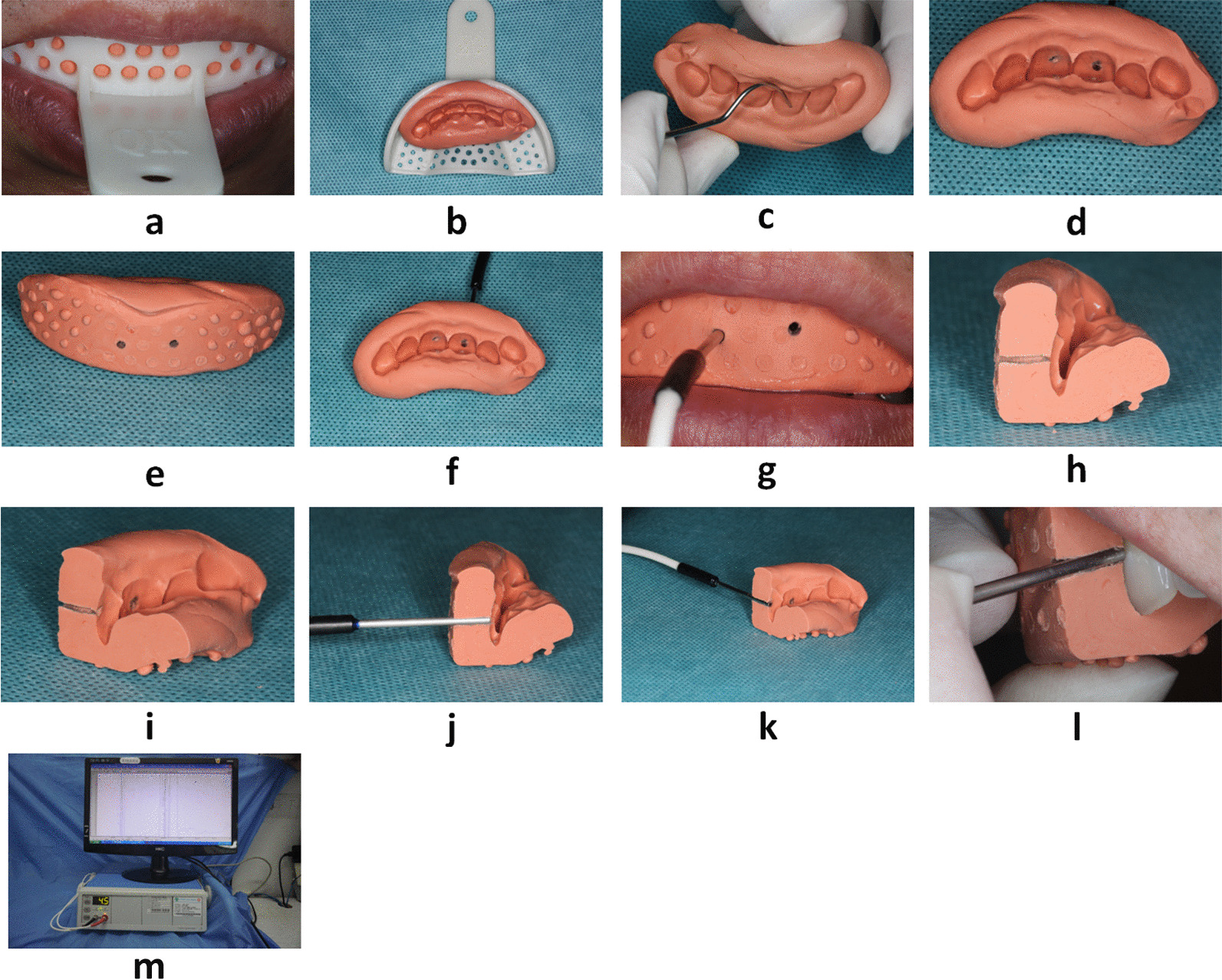
Fig. 1.

Presentation of the detection process that used a doppler blood flow tester: a preparation of silicone rubber impression of upper anterior teeth of patients that used heavy body impression putty; b silicone rubber impression preparation removed from the oral cavity and disinfected; c marking the anatomical location for probing; (d and e) lingual and labial views after drilling the holes; f test probe trimming and punching position; g placement of the test probe; h longitudinal section of silicon rubber offset printing die after cutting; i view of the lingual surface of the longitudinal section after the silicon rubber offset printing die is cut; (j and k) probe placement after longitudinal sectioning the silicon rubber offset die; (l) placement of the probe in the sectioned silicone rubber impression when testing in the oral cavity; (m) LDF equipment in the clinical settings
