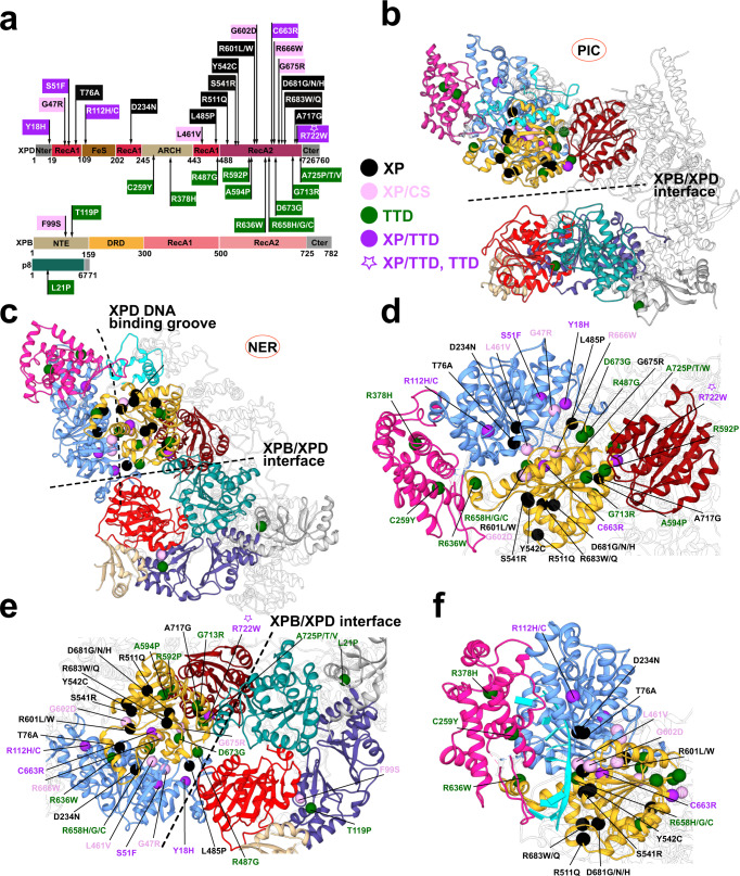
Fig. 7. Human disease mutations mapped onto TFIIH show distinct patterns within protein-protein and community interfaces.
a TTD, XP, XP/CS and XP/TTD point mutations mapped onto XPD, XPB, and p8 subunits do not co-localize by disease on primary sequence. b Map of human disease mutations (spheres) onto the PIC-TFIIH structure shown in cartoon representation and colored by community. c Map of human disease mutations (spheres) onto the NER-TFIIH structure shown in cartoon representation and colored by community. d Zoomed view of mutations within XPD. e Zoomed view of mutations at the XPB–XPD interface. f Zoomed view of mutations lining the path of ssDNA in XPD.

