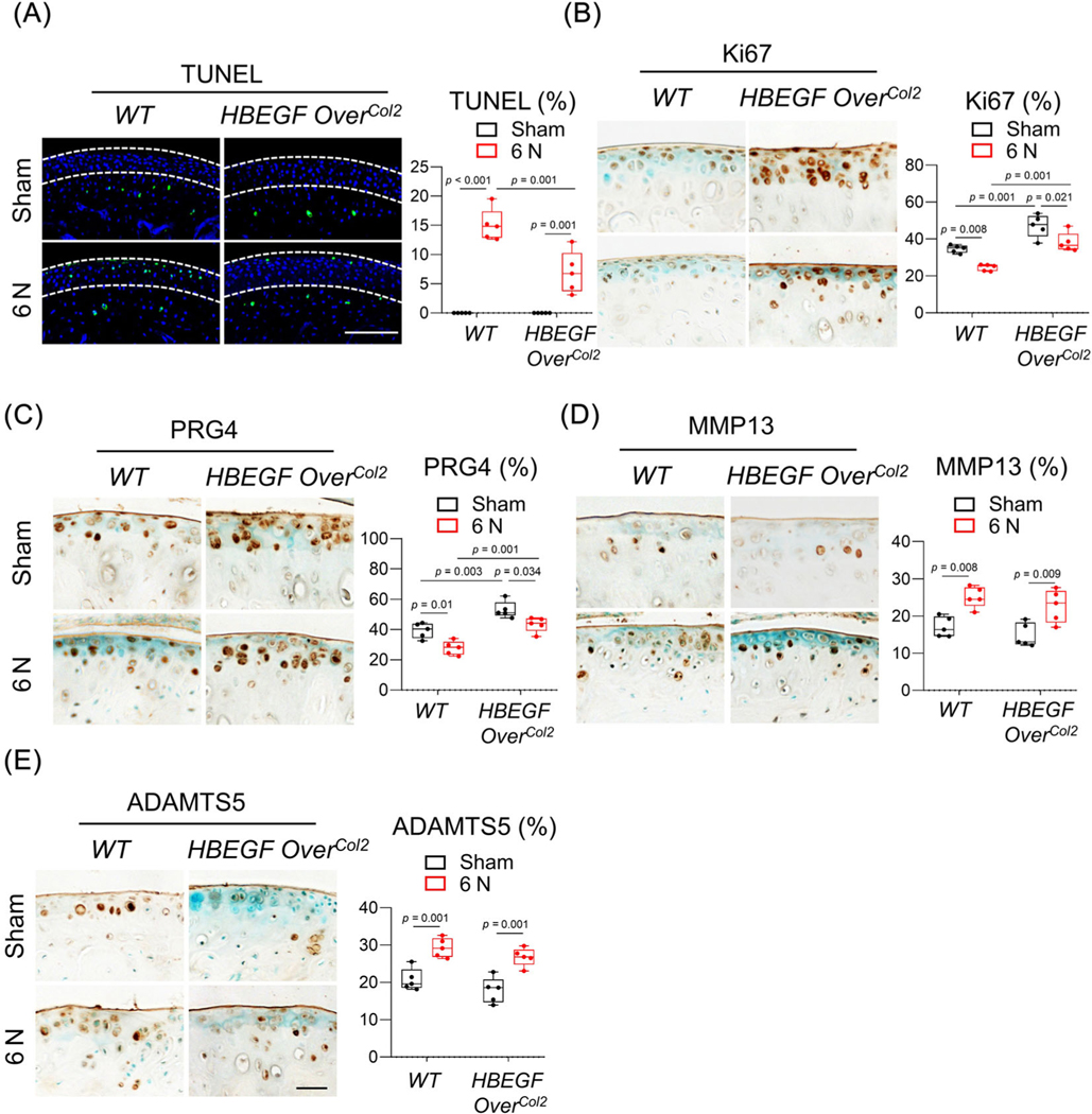
Fig. 4.
EGFR signaling promotes the survival, proliferation, and lubrication of chondrocytes in articular cartilage. (A) TUNEL staining of WT and HBEGF OverCol2 articular cartilage at 4 days after 6 N loading. White dashed lines in the images outline uncalcified zone. The percentage of TUNEL+ cells in uncalcified cartilage was quantified and is shown at the right (n = 5/group). Scale bars, 100 μm. (B–E) Ki67 (B), PRG4 (C), MMP13 (D), and ADAMTS5 (E) staining of WT and HBEGF OverCol2 articular cartilage at 2 weeks after 6 N loading. In each panel, the percentage of positive cells in the articular cartilage is quantified at the right (n = 5/group). Scale bars, 50 μm. The comparisons were conducted by two-way ANOVA with Tukey–Kramer multiple comparison test.

