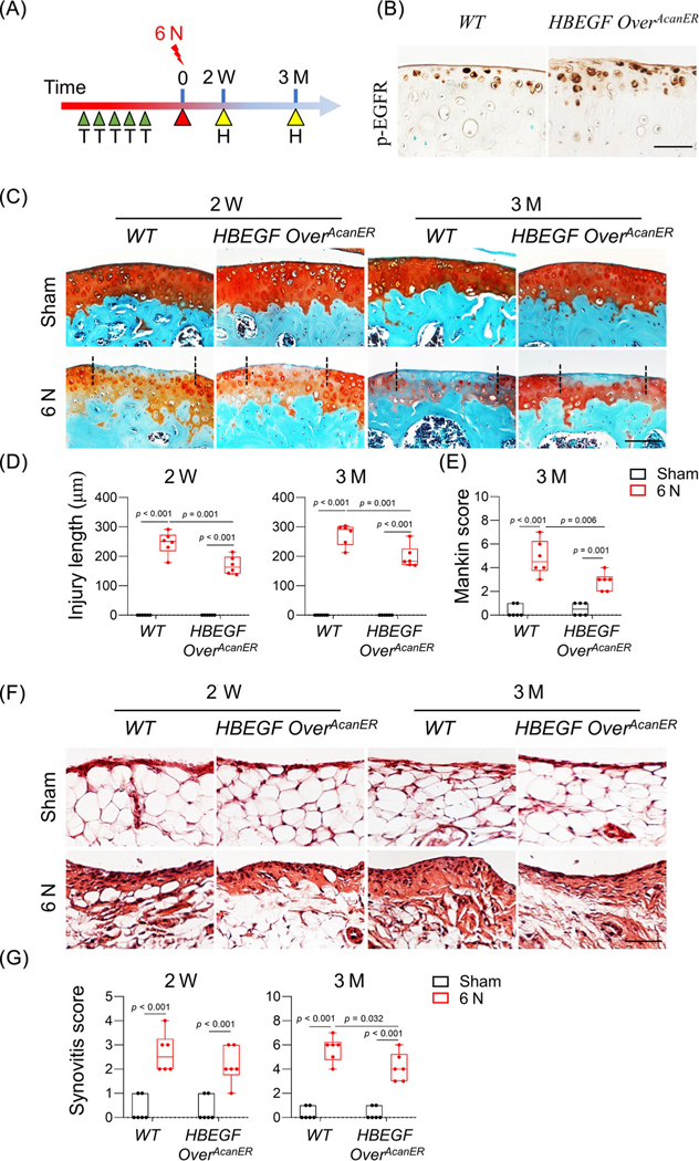
Fig. 5.
Chondrocyte-specific overactivation of EGFR signaling in adult mice hinders PTOA progression. (A) Study design showing WT and HBEGF OverAcanER mice received tamoxifen injections right before 6 N loading. Their knee joints were harvested 2 weeks and 3 months later. H, harvest; T, tamoxifen injection. (B) Immunostaining of p-EGFR in uninjured tibial articular cartilage from WT and HBEGF OverAcanER mice at 2 weeks after loading. Scale bars, 50 μm. (C) safranin-O/Fast green-stained sections of WT and HBEGF OverAcanER cartilage at indicated time points. Scale bars, 100 μm. (D) Cartilage injury length was quantified (n = 6/group). (E) Mankin score of WT and HBEGF OverAcanER mouse joints at 3 months after loading was quantified (n = 6/group). (F) H&E-stained sections of WT and HBEGF OverAcanER synovium at indicated time points. Scale bars, 50 μm. (G) Synovitis score was quantified (n = 6/group). The comparisons were conducted by two-way ANOVA with Tukey–Kramer multiple comparison test.

