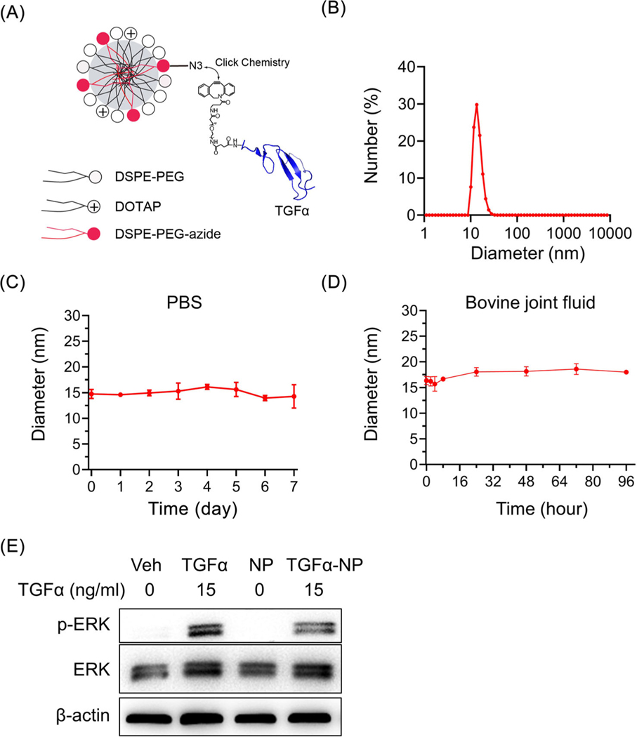
Fig. 7.
Synthesis and characterization of novel nanoparticle conjugated with TGFα. (A) Schematic diagram of TGFα-NP. (B) Size distribution of TGFα-NP as evidenced by DLS. (C) The diameter of TGFα-NP was measured in PBS for up to 7 days. (D) The diameter of TGFα-NP was measured in bovine synovial fluid for up to 96 h. (E) Western blot of p-ERK, an EGFR downstream target, in primary chondrocytes at 15 min after TGFα-NP (15 ng/ml of TGFα content) treatment. Cells treated by vehicle (PBS), free TGFα (15 ng/ml), and empty-NP (i.e., no TGFα conjugation) were used as controls. The comparisons were conducted by ordinary one-way ANOVA with Tukey–Kramer multiple comparison test.

