Fig. 2.

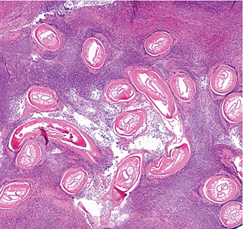
Microscopic slide with hematoxylin and eosin stain demonstrating multiple sections of filarial nematodes, surrounded by eosinophil granulocytes.

Microscopic slide with hematoxylin and eosin stain demonstrating multiple sections of filarial nematodes, surrounded by eosinophil granulocytes.