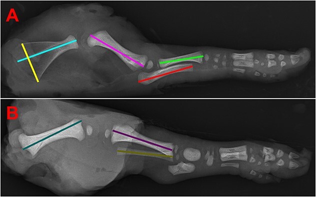
Figure 1.

Images of fetal morphometric and forelimb and hindlimb bone measurements. (A) Radiograph of fetal forelimb used to measure width of scapula (yellow), length of scapula (light blue), humerus (pink), ulna (red), and radius (green). Long bones were measured for length by using a straight line through the median record the distance from the proximal ends to the distal ends. Scapula width was measured by a straight line that ran from the tip of the inferior angle to the tip of the superior angle. The length of the scapula was recorded by a straight line dissecting the bone along the central ridge. (B) Radiograph of fetal hindlimb used to measure length of femur (teal), tibia (purple), and fibula (gold).
