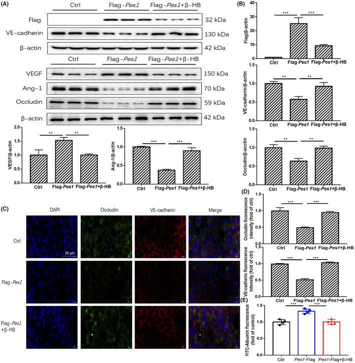
FIGURE 6.

β‐HB treatment impaired the increment of paracellular permeability by in vitro supplementation of Pes1. (A, B) The protein levels of PES1, VEGF, VE‐cadherin, Ang‐1 and Occludin in MVECs were detected by immunoblotting after Flag‐Pes1 plus β‐HB treatment. (C, D) Shown are immunofluorescence images of Flag‐Pes1 plus β‐HB‐treated MVECs for Occludin and VE‐cadherin expression and localizations, scale bar represents 20 μm. The nuclei were stained with DAPI. (E) Exhibited is the paracellular permeability in the cultured MVECs in different groups. Data were represented as mean ± SEM, each experiment was performed independently three times. **p < 0.01, ***p < 0.001 compared with control (anova, Student–Newman–Keuls q‐test).
