Fig. 1.
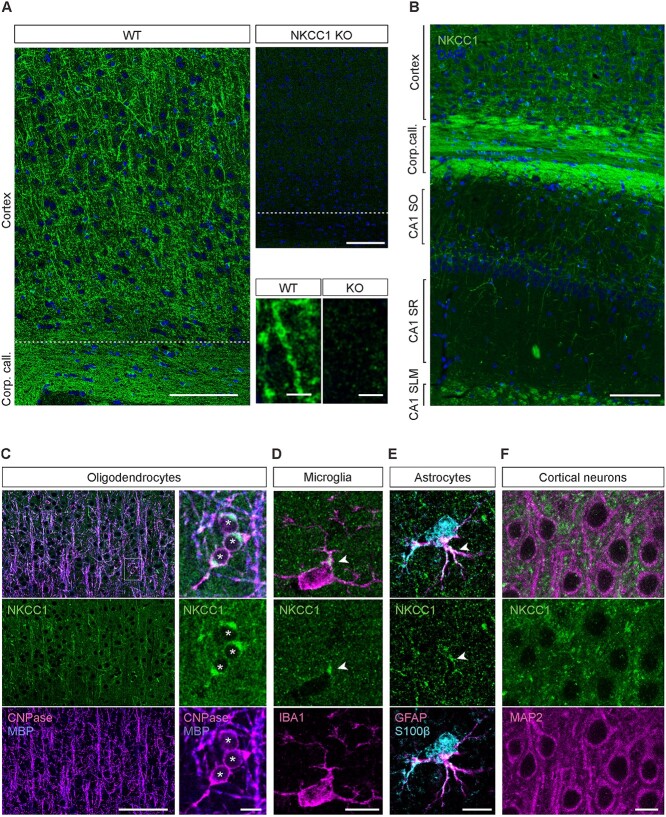
NKCC1 protein is strongly expressed in glial cells of the adult mouse cortex and hippocampus. (A) NKCC1 immunoreactivity in the somatosensory cortex of WT mice. Sections from NKCC1 KO mice show a weak and homogenous background signal. Identical results were obtained with both GpA and RbC NKCC1 antibodies (n = 4 WT/KO pairs). Higher magnification images are shown in the bottom right corner. (B) Overview of the NKCC1 expression in the cortex and hippocampus. (C) The strong cortical and callosal NKCC1 protein signal mostly originates from oligodendrocytes, colocalizing with MBP and the oligodendrocytic marker CNPase. (D) Microglial cells, detected by their Iba1 expression, showed one or few clusters of NKCC1 IR in the soma, often close to a ramification (arrowhead). (E) Astrocytes, identified by expression of both GFAP and S100B, showed NKCC1 expression in some of their rami (arrowheads). (F) Cortical neurons identified by MAP2 IR do not express detectable levels of NKCC1 in the somatodendritic compartment. Scale bars A: 100 μm (top row pair) and 5 μm (bottom row pair), B: 100 μm, C: 100 μm (left) and 10 μm (right), D–F: 10 μm.

