Fig. 5.
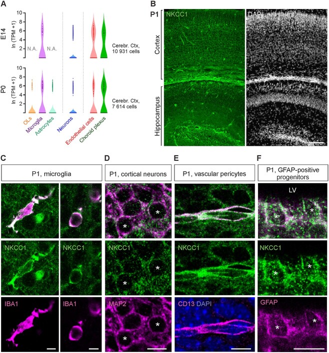
In the P1 cortex and hippocampus, NKCC1 is strongly expressed in microglia, blood vessels, progenitor cells, and neurons. (A) scRNA-seq analysis indicates expression of NKCC1 at E14 and P0 in vascular endothelial cells, choroid plexus endothelium, microglia, and neurons, and at P0 also in astrocytes and oligodendrocytes. Data extracted from (Loo et al. 2019). (B) Overview of the NKCC1 IR in the P1 cortex and hippocampus. (C) Strong NKCC1 IR is visible in both ramified (left panel) and ameboid (right panel) microglia. (D) Clear NKCC1 IR was found on cortical and hippocampal pyramidal neurons. (E) At P1, blood vessels were strongly immunoreactive for NKCC1, with the signal colocalizing with the pericytic marker CD13. (F) GFAP-positive cells close to ventricular wall displayed robust NKCC1 IR. LV = Lateral ventricle. Scale bars B: 100 μm, C: 5 μm, D–F: 10 μm.

