. 2023 May 15;13(21):14685–14697. doi: 10.1039/d3ra02092d

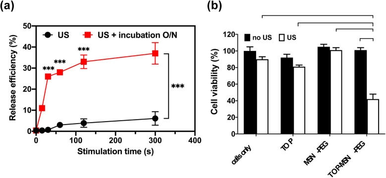
Fig. 4. Ultrasound-stimulated TOP release in RPMI medium using a probe sonicator. (a) Time-dependent release profile of TOP from MSN-PEG immediately (black) or after a 20 h incubation (red) post probe sonication. (b) Cell viability of OVCAR-3 cells treated with TOP-MSN-PEG, the equivalent amount of TOP alone (positive control), MSN-PEG, and cells only (negative control), with or without ultrasound stimulation (power density: 75 W cm2; stimulation time: 30 s). Cell viability was determined by the CCK-8 assay and normalized to the control group without any treatment. Data are means ± SD of three independent experiments (***P < 0.001).

Fig. 4