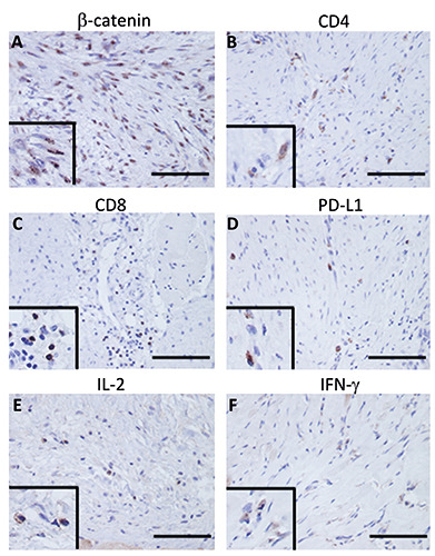
Figure 1.

Representative immunostaining for each immune molecule. β-catenin showed staining primarily in the cytoplasm of the tumor cells (A). CD4 (B) and CD8 (C) showed staining primarily in tumor-infiltrating lymphocytes. PD-L1 showed staining mainly in DT cells (D). IL-2 (E) and IFN-g (F) were found to be stained in tumor cells and lymphocytes; scale bar: 100 μm. The wipe screens show a higher magnification field.
