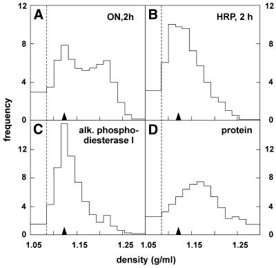
Figure 4.
Density distribution after 125I-ON pulse. HepG2 cells were incubated at 37°C for 2 h with either 125–250 nM 125I-ON (A) or 100 µM HRP (B), washed and surface digested with pronase at 4°C. After homogenisation, postnuclear particles were loaded on the top of linear sucrose density gradients (loading zone is left of the dotted line) and centrifuged to equilibrium. Distributions were normalised as density/frequency histograms. Data shown are averages of three fractionation experiments and compared with the distribution of the plasma membrane/endosomal marker, alkaline phosphodiesterase I (C), and of total particulate protein (D). Recoveries ranged between 89 and 114%. For convenience, the position of ON low-density peak (1.13 g/ml in density; filled arrowheads) is repeated on each distribution.

