Table 3.
Imaging Findings or Classifications Useful for Indicating Procedures to Treat MRCTs a
| Finding/Classification | Use/Correlations | |
|---|---|---|
| Radiograph | ||
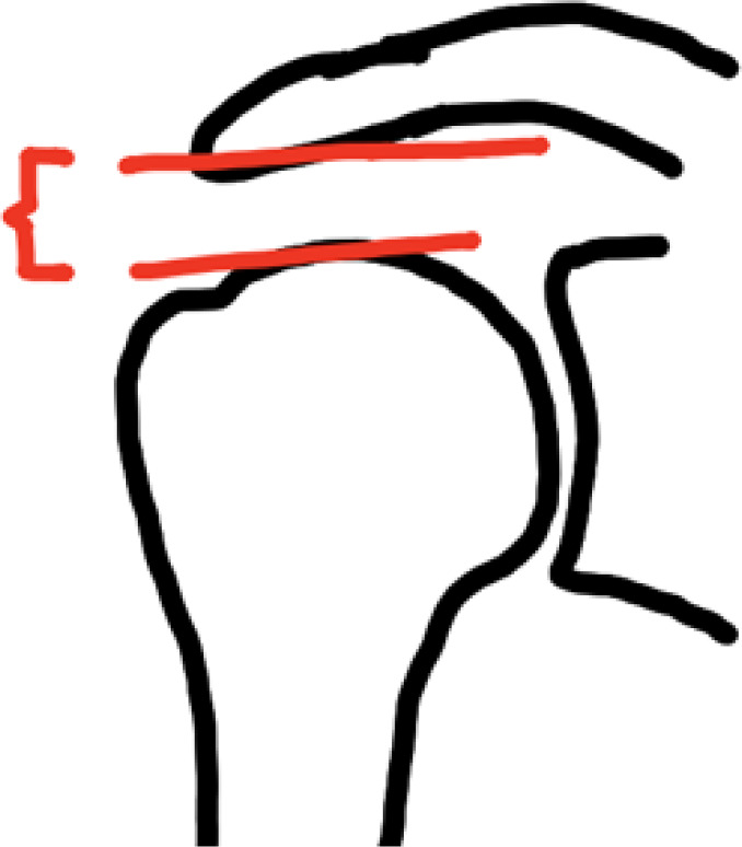
| Acromiohumeral interval |
|
Indicator of vertical force couple imbalance. Stress examination can assess static versus dynamic superior migration. 40 |
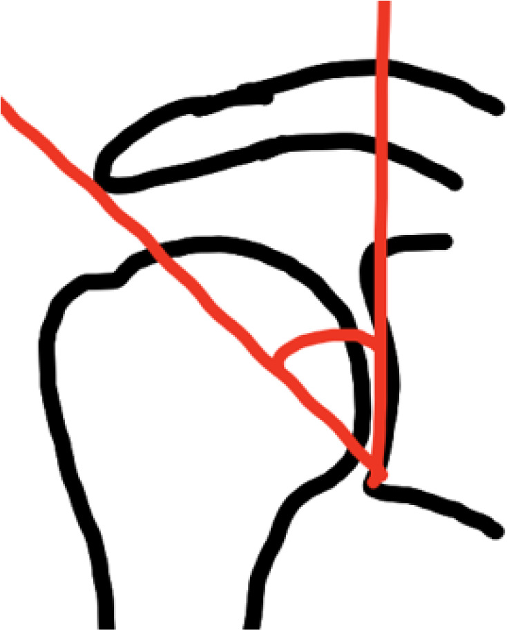
| Critical shoulder angle |
|
Describes how vertical the moment arm of the deltoid is, and how much the shoulder relies on the cuff for initiation of overhead motion.28,60 |
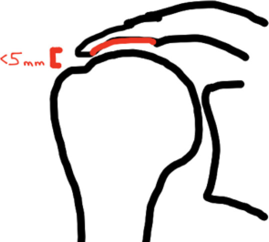
| Hamada classification |
|
Describes radiographic progression, from muscle imbalance causing proximal migration, to bone remodeling of the acromion, and finally glenohumeral arthritis. 34 Joint-preserving procedures with generally inferior outcomes for Hamada ≥3. 27 |
| MRI | ||
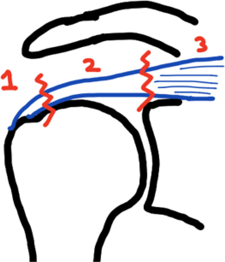
| Patte |
|
Describes extent of tendon retraction. 74 |
| Tangent sign |
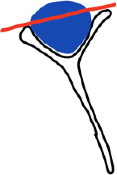
|
Describes extent of muscle atrophy.42,97 |
| Goutallier | 1 = normal muscle 2 = fatty streaks 3 = muscle > fat 4 = fat > muscle |
Describes extent of muscle fatty infiltration. 33 |
| Tendon stump length |
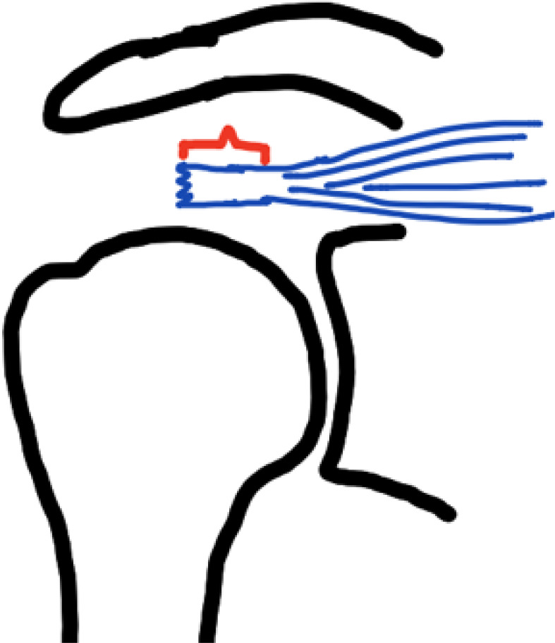
|
<15 mm correlates with 92% repair failure rate. 56 |
a MRCT, massive rotator cuff tear; MRI, magnetic resonance imaging.
