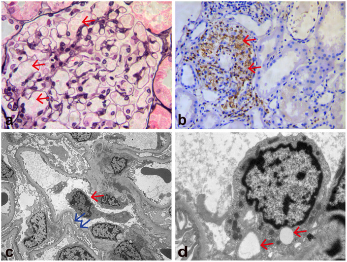
Figure 1.
Kidney biopsy findings. (a) Light microscopy shows that the glomeruli are enlarged and the glomerular capillary loops are filled with a large number of foamy cells (red arrows). The endothelial cells are swollen and show vacuolar degeneration (periodic acid-methenamine silver staining; original magnification, ×400). (b) Light microscopy shows granular cytoplasmic staining (CD68 immunohistochemical stain) of abundant intracapillary infiltrating histiocytes and a few interstitial histiocytes (red arrows; original magnification, ×200) and (c, d) Electron microscopy shows macrophages in the glomerulus (red arrow in c), effacement of most foot processes (blue arrows in c), endothelial swelling, and vacuolar degeneration (red arrows in d). No hemophagocytosis or immune-type electron dense deposits were observed (original magnification, c: ×4000; d: ×15,000).

