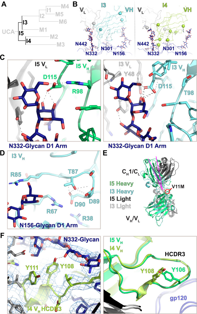
Fig. 3. The I5 to I3 and I4 path split.
A DH270 clonal tree highlighting the I5, I3, and I4 intermediate antibodies. B Top views of the I3 and I4 contact sites. Alpha-carbons (Cα) of heavy and light chain mutations are presented as spheres. C (left) The N332-glycan D1 arm contact with the I5 VH/VL cleft. (right) The N332-glycan D1 arm contact with the I3 VH/VL cleft. D Structure of the I3 intermediate at the R87T site near potential N156-glycan contacts. E Aligned I5 and I3 VH domains highlighting the change in elbow angle (magenta). F (left) Map and fit coordinates of the new N332-glycan contact formed at the I4 HCDR3 S108Y mutation site. (right) Aligned I5 and I4 VH domains showing HCDR3 loop arrangements.

