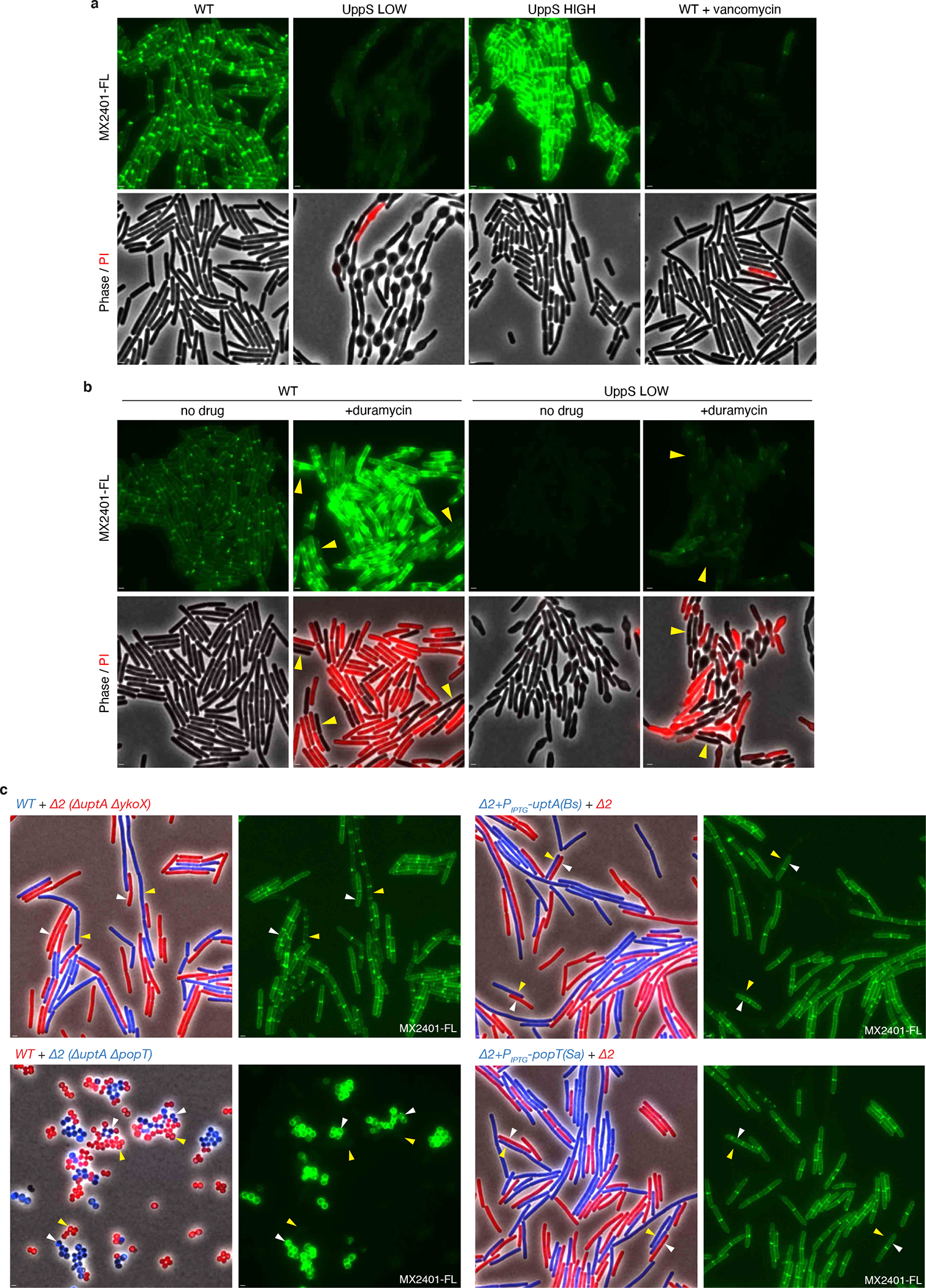
Extended Data Fig. 7. MX2401-FL specifically labels outward-facing UndP.

(a) Validation of MX2401 conjugated to CF488 (MX2401-FL). Representative fluorescence and phase-contrast images of the indicated strains labeled with MX2401-FL and propidium iodide (PI). Cells with reduced de novo synthesis of UndP (UppS LOW) harbor an IPTG-regulated allele of uppS (Pspank-uppS) and were propagated in the presence of 4 μM IPTG. Cells with increased de novo synthesis of UndP (UppS HIGH) harbor a stronger IPTG-regulated allele of uppS (Physpank-uppS) and were propagated with 500 μM IPTG. UppS LOW cells have low MX2401-FL staining and bulge due to impaired for cell wall synthesis. UppS HIGH cells have high MX2401-FL staining and are shorter. Cells treated with vancomycin for 5 minutes prior to MX2401-FL staining trap UndP in lipid II and have low MX2401-FL signal. All MX2401-FL images were normalized identically with minimum and maximum intensities of 125 and 600 to detect weak MX2401-FL staining in the UppS LOW and vancomycin-treated strains. (b) Wild-type and UppS LOW strains were stained with MX2401-FL or a mixture of MX2401-FL and duramycin, which generates pores in the membrane allowing MX2401-FL access to the cytoplasmic-facing UndP. Cells with membrane permeability defects as assayed by PI have higher MX2401-FL signal. Cells with intact membranes (yellow carets) have lower MX2401-FL staining. All MX2401-FL images were normalized identically with minimum and maximum intensities of 125 and 1500 to prevent saturating the MX2401-FL signal. (c) Representative microscopy images of the indicated strains. Strains expressing different fluorescent proteins (B. subtilis) or labeled with different fluorescent D-amino acids (S. aureus) were mixed and then stained with MX2401-FL. (Left) overlays of phase contrast and fluorescent images in the red and blue channels to distinguish the two strains. (Right) MX2401-FL staining. Yellow carets highlight wild-type cells or cells over-expressing UptA(Bs) or PopT(Sa). White carets highlight cells lacking the UndP transporters. Scale bar, 1 μm.
