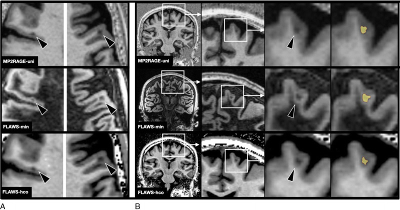
FIGURE 2.

A, Example of 2 cortical lesions (arrowheads) on MP2RAGEuni (top row), FLAWSmin (mid row), and FLAWShco (bottom row). B, “Zoom in” on a cortical lesion on MP2RAGEuni (top row), FLAWSmin (mid row), and FLAWShco (bottom row) and its segmentation (in yellow). Note that the clear delineation on FLAWSmin allows a slightly larger segmentation of the cortical lesion. FLAWS indicates fluid and white matter suppression; MP2RAGE, magnetization prepared 2 rapid acquisition gradient echo.
