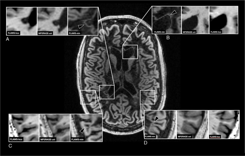
FIGURE 5.

FLAWSmin of a patient with MS demonstrating the “halo” appearance. Partial volume effects at the border of tissues with high divergence of signal intensities lead to a hyperintense band at the respective border. MP2RAGEuni and FLAWShco are shown for reference. A, Hyperintense band (arrowhead) around a periventricular white matter lesion (not systematically assessed in this study). B, Hyperintense band (arrowhead) at the normal-appearing border between CSF and brain tissue. C, Hyperintense band (arrowhead “halo”) around a leukocortical lesion. D, Hyperintense band (arrowhead, “halo”) around an intracortical lesion. FLAWS indicates fluid and white matter suppression; MP2RAGE, magnetization prepared 2 rapid acquisition gradient echo.
