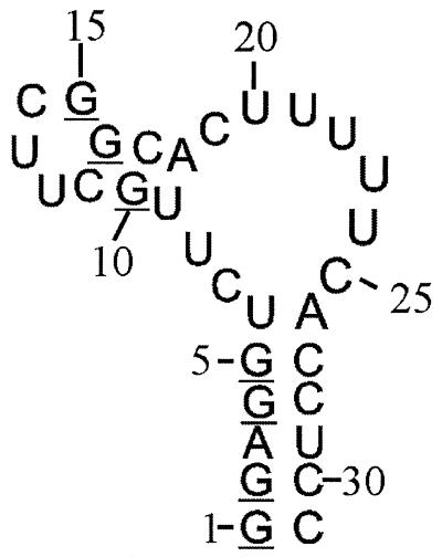
Figure 1.
Secondary structure of the PBS of hepatitis B virus, synthesised to illustrate the stereo-specific labelling method. In one sample the G residues have 13C labels in the ribose ring, deuterium labels at 1′, 3′ and 4′ positions and stereo-specifically at the 5′′ position, the C and U residues have deuterium labels at the 1′, 3′, 4′, 5′ and 5′′ positions, and the A residues are fully protonated and not enriched in 13C or 15N. The stereo-specifically 5′′-deuterated G residues are underlined. In a second sample all residues were uniformly 13C/15N-labelled.

