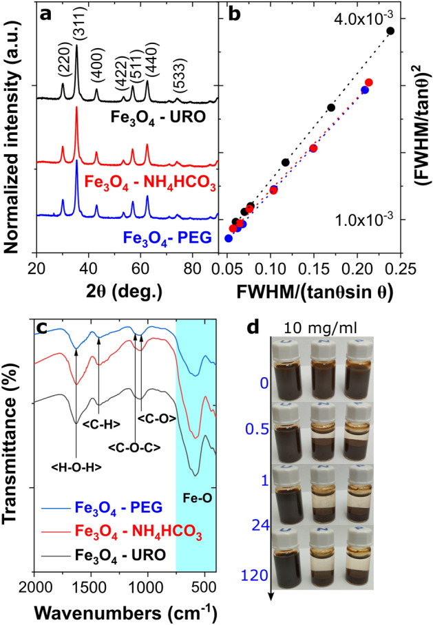
Figure 2.

(a) XRD patterns of synthesized magnetite nanoparticles with characteristic Miller indices (Fd-3 m space group; 00–019-0629 card number), (b) Halder-Wagner plots from (220), (311), (400), (422), (440) and (511) diffraction peaks (R2 above 0.99 in all cases), (c) FTIR spectra of magnetite nanoparticles with marked identified vibrations related to the presence of the functionalized surface, (d) macroscopic images of the changes in the stability of colloidal dispersion of magnetite nanoparticles with high 10 mg/ml concentration in the time domain, i.e. from 0 to 120 h (from left: Fe3O4—URO NPs, Fe3O4—NH4HCO3 NPs and Fe3O4—PEG NPs).
