
Figure 3.
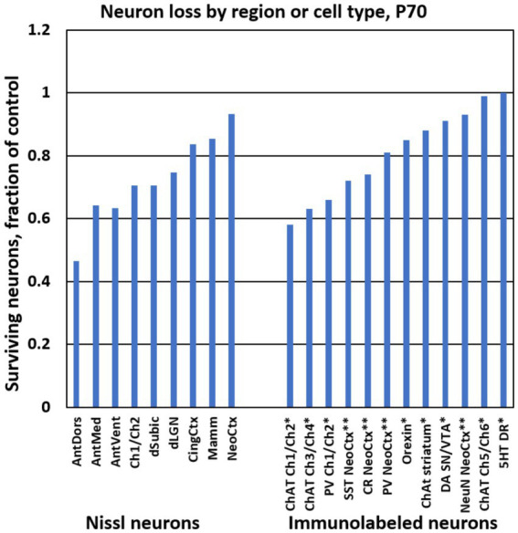
Bar graphs show the ethanol-induced neuron loss at P70 from different brain regions in the present study (“Nissl neurons,” at left). For comparison, at right are stereological estimates of immunolabeled neuron populations from previous studies using the same treatment paradigm. * Results are from Smiley et al. (2021). ** Results are from Smiley et al. (2019). Abbreviations: 5HT DR, serotonin neurons of dorsal raphe; AntDors, anterior dorsal thalamic nucleus; AntMed, anterior medial thalamic nucleus; AntVent, anterior ventral thalamic nucleus; ChAT, choline acetyl transferase immunolabeled cells; Ch1/Ch2, medial septum and vertical diagonal band region; Ch3/Ch4, horizontal diagonal band and nucleus basalis region; Ch5/Ch6, pedunculopontine and lateral dorsal tegmental nuclei; CingCtx, cingulate cortex; CR, calretinin immunolabeled cells; DA SN/VTA, dopamine cells of substantia nigra and ventral tegmental area; dLGN, dorsal lateral geniculate nucleus; dSubic, dorsal subiculum; Mamm, mamillary bodies; NeoCtx, neocortex; NeuN, neuronal nuclear antibody immunolabeled cells; Orexin, orexin cells of the hypothalamus; PV, parvalbumin immunolabeled cells; SST, somatostatin immunolabeled cells.