. 2023 May 15;24(5):455–457. doi: 10.1631/jzus.B2300026

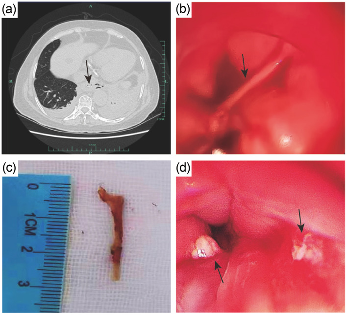
Fig. 1. Images of the esophageal foreign body. (a) The chest computed tomography (CT) suggested a foreign body at the end of the esophagus (indicated by the black arrow). Gas accumulation on the left side and perforation was found, along with bilateral pneumonia, bilateral pleural effusion, and partial distension of the left lower lobe. (b) A foreign body (indicated by the black arrow) was inserted into the wall of the esophagus at 35 cm of the esophagus from the incisor, and produced pus emission. (c) The esophageal foreign body: a duck bone approximately 3 cm in length. (d) The mucosa of the upper esophagus was swollen after removal of the foreign body.

Fig. 1