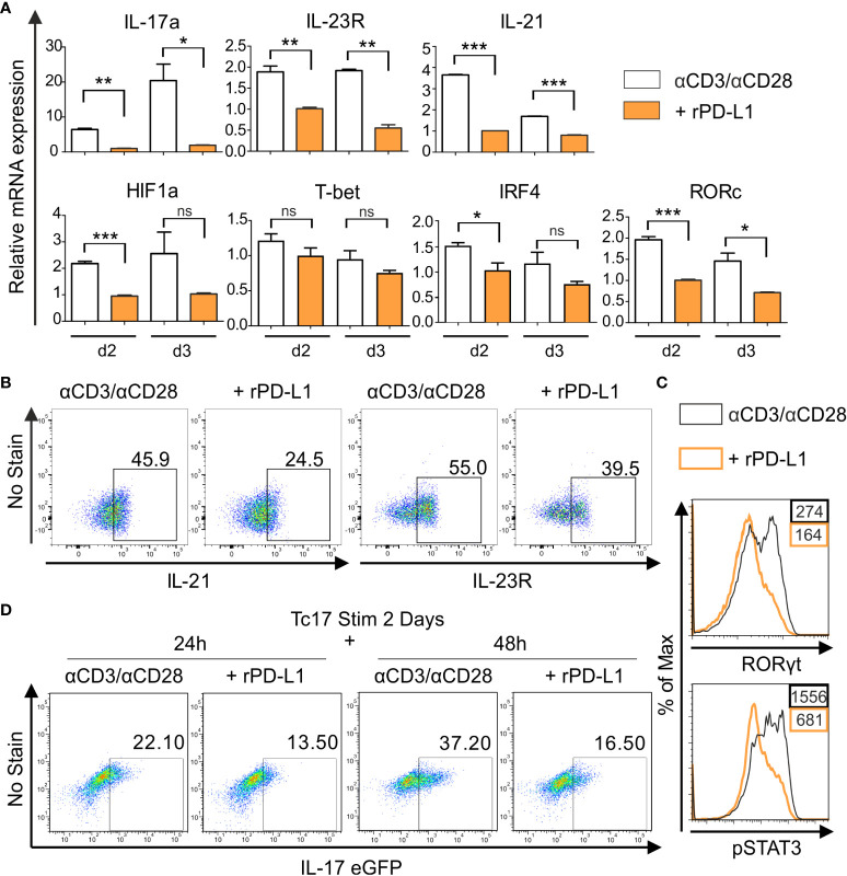
Figure 3.
PD-1 suppresses Tc17 hallmarks. (A) CD8+ T-cells from C57BL/6 mice were stimulated as in (2C) and harvested at indicated time points. The harvested cells were lysed, RNA was extracted, and RT was used to synthesize cDNA. The relative expression levels of the indicated genes analyzed using real-time PCR are shown as mean+SEM of replicates from an experiment. (B, C) Tc17 cells were stimulated as in (A) and expression of IL-21 and IL-23R (B), RORγt and pSTAT3 (C) was measured by flow cytometry 3 days after activation. (D) CD8+ T-cells from IL-17A-eGFP reporter mice were stimulated with microspheres immobilized with anti-CD3, anti-CD28 for 2 days under Tc17 conditions. These cells were then engaged with microspheres immobilized with rPD-L1 or not, associated with anti-CD3 and anti-CD28 for indicated time points and thereafter IL-17 expression was measured by flow cytometry. The data is representative of two to three independent experiments. ***P<0.001, **P<0.01, *P<0.05; ns, not significant, calculated by unpaired t-test.

