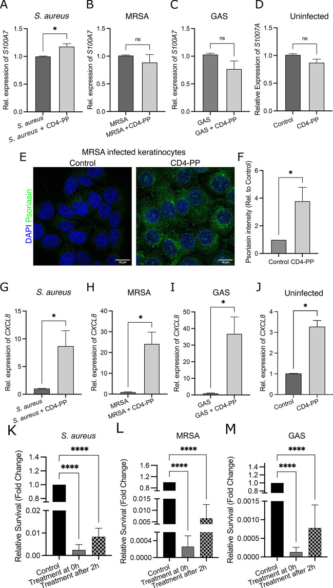
Figure 2.
Increased psoriasin and CXCL8 expression in keratinocytes stimulated with CD4-PP contributes to a decrease in infection. (A) During S. aureus infection, keratinocytes showed an increase in S100A7 levels when treated with CD4-PP (*p < 0.05, paired t test). No differences in S100A7 expression were observed in (B) MRSA- or (C) GAS-infected keratinocytes nor in (D) uninfected keratinocytes treated with CD4-PP. (E) Representative images of keratinocytes infected with MRSA with or without CD4-PP treatment depicting psoriasin peptide (green) and nucleus (blue). (F) Densitometric analysis of psoriasin staining in MRSA-infected samples showed a significant increase of keratinocytes treated with CD4-PP (*p < 0.05, unpaired t test). CD4-PP increased the expression of CXCL8 in keratinocytes infected with (G) S. aureus (ATCC 29213), (H) MRSA (CCUG 31966), and (I) GAS (ATCC 19615) and (J) uninfected keratinocytes (*p < 0.05, paired t test). Survival of skin pathogens is shown for (K) S. aureus (ATCC 29213), (L) MRSA (CCUG 31966), and (M) GAS (ATCC 19615) after infecting keratinocytes. The survival in treatment groups is relative to untreated control. CD4-PP was initiated at the same time as infection (treatment at 0 h) or after 2 h of infection (treatment after 2 h) (****p < 0.0001, one-way ANOVA). Relative mRNA expression of target genes was performed in at least three independent sets in duplicate or triplicate. Microscopy imaging and densitometry analysis were performed in three independent experiments, with each experiment consisting of 4–5 random view fields. The average integrated density of each cell per set used for statistical analyses.

