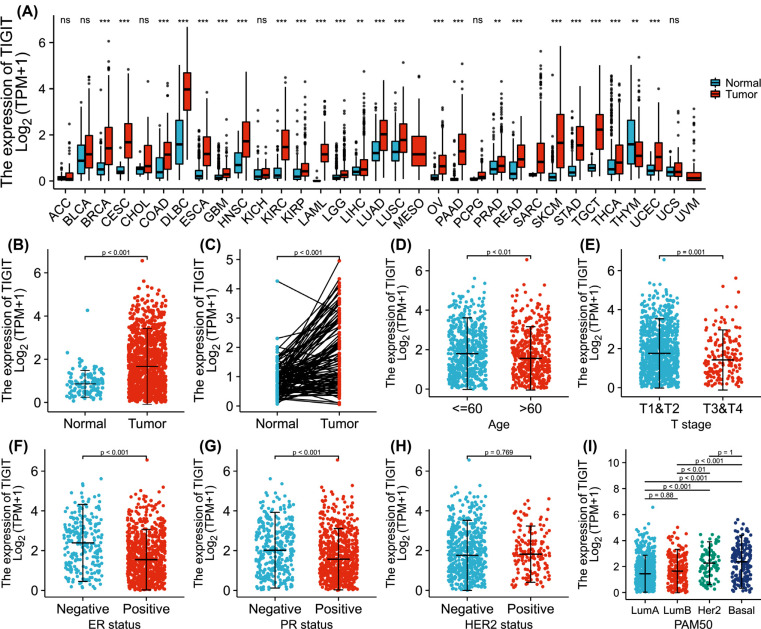
Fig. (1).
The levels of TIGIT in cancer. (A) relative to corresponding normal tissues, the level of TIGIT in different cancer tissues is different (*p<0.05); (B) The levels of TIGIT in invasive breast cancer were markedly high than in normal tissues (p<0.001); (C) In the paired samples, the levels of TIGIT in invasive breast cancer was significantly increased (p<0.001); (D-I) Association between TIGIT and clinical manifestations of invasive breast cancer.

