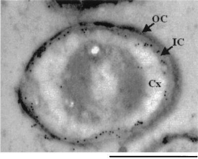
FIG. 3.
Localization of SafA in wild-type spores. Immunogold electron microscopy was used to localize SafA in spores. Electron-dense gold particles indicate the position of anti-SafA in the electron micrograph of a wild-type spore. Few or no gold particles decorated SafA mutant spores (data not shown). The spore cortex (Cx), outer coat (OC), and inner coat (IC) are indicated. Bar, 500 nm.

