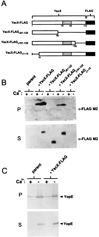
FIG. 4.
Secretion of YscX-FLAG and truncated YscX-FLAG proteins from the parent strain Y. pestis KIM8-3002. (A) Schematic representation of FLAG-tagged YscX (YscX-FLAG), YscX-FLAGΔ61–122, YscX-FLAGΔ101–122, and YscX-FLAGΔ1–15. A predicted coiled-coil domain spanning residues 70 to 93 of YscX is shown (hatched box). (B) Immunoblot analysis of SDS-PAGE-separated culture supernatant (S) and cell pellet (P) fractions from Y. pestis KIM8-3002 (parent) and KIM8-3002 transformed with plasmids pFLAG-YscX, pFLAG-YscXΔ61–122, pFLAG-YscXΔ101–122, and pFLAG-YscXΔ1–15. The FLAG M2 monoclonal antibody was used to detect the FLAG-tagged products. (C) Immunoblot analysis of SDS-PAGE-separated culture supernatant (S) and cell pellet (P) fractions from Y. pestis KIM8-3002 (parent) and KIM8-3002 complemented with plasmid pFLAG-YscX. Antiserum specific for YopE was used to detect this protein in the supernatant and cell pellet fractions.

